Science time! Today we’re going to talk about extracellular vesicles. You know, those things that dude down the corridor works on that you don’t really believe are a thing.
We’ll go the semi-patronizing route I took with the stroke chat and I’ll tell you a little bit about the history of the field, a little bit about what extracellular vesicles are, and a little bit about why they’re interesting.
We’re going to start in the 40s, where you could get away with writing a series of 30 papers on basically the same thing, each with only two beautifully hand drawn graphs and one meticulous table in. There were these two chaps, Chargaff and West who were a biochemist and a medic, respectively. They were trying to figure out how all the disparate components of blood came together to make a clot by spinning down and separating all the components to see what they did. In 1936 they began a series of papers called ‘Studies on the Chemistry of Blood Coagulation’ and paper 19 of the series is what we think might be the start of the field of extracellular vesicle research. They found that “the addition of the high speed sediment to the supernatant plasma brought about a very considerable shortening of the clotting time”. Basically, in the truly broadest sense (don’t hate me extracellular vesicle people) if you spin down blood with anticoagulant in it you get plasma, if you spin down that plasma you get extracellular vesicles (and a load of other stuff but we’ll get into that on a later occasion).

Erwin Chargaff, Studies on the Chemistry of Blood Coagulation, https://doi.org/10.1016/S0021-9258(18)73959-8
In the next paper in the series they took plasma from a normal adult, a haemophiliac and a female with what is described as a ‘clotting disorder’ with no apparent interest in which one. You can tell this is the 30s because the details are sparse and the n is 3. They timed how long it took for clots to form in all of these patients and found, no surprise, the haemophiliac and the female with the clotting disorder didn’t clot all that quickly. They then took the sediment they found earlier, the one I said is probably the extracellular vesicle fraction, and applied it to these patients’ bloods and found that the time to clot was much quicker. Whilst they said at the time that this fraction “probably includes, in addition to the thromboplastic agent, a variety of minute breakdown products of blood corpuscles” they did not call them extracellular vesicles or explore them any further.
Now, between the 40s and the 80s there is a ton of literature that never really went anywhere. People seem to have found extracellular vesicles in all sorts of places and not pursued them as anything exciting or novel. If you’re interested and patient there’s a history of extracellular vesicle research review due out sometime soon, care of yours truly and a whole bunch of much more eminent extracellular vesicle people. But where the field really started to come into its own was in the 1980s.
Rose Johnstone and Philip Stahl were looking at receptor trafficking. They both published papers in 1983 on the transferrin receptor. In these papers they had found the receptor on vesicles within multivesicular bodies and had found these vesicles were released by exocytosis. Whilst the term ‘exosome’ cannot be attributed to either Johnstone or Stahl, but rather to Eberhard Trams of Ursula Heine’s lab, Rose Johnstone was the first to officially describe exosomes. She hailed them as “lipoprotein structures with a phospholipid composition characteristic of red cell membranes”.
This leads us neatly to modern classification systems and the sticky mess therein. Most people will have heard of exosomes but it’s now considered a rather old-school term and the more widely accepted one is extracellular vesicles, or EVs as I will now call them because it’s less of a mouthful. EVs can be broadly classified into two camps according to their biogenesis; large EVs (usually more than 150nm) are shed from the cell surface, these used to be called microvesicles or microparticles, small EVs (usually less than 150nm) are formed by invagination of the membrane, they’re stored in the multivesicular body and released by exocytosis, these used to be called exosomes. The problem here is there is a lot of overlap in sizes and some shared markers between the different classes. ISEV, the International Society for Extracellular Vesicles, has published some great position papers on this issue so feel free to go and peruse should you wish to know more.
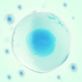
Extracellular vesicles (EVs) are lipid bound vesicles secreted by cells into the extracellular space. The three main subtypes are microvesicles (MVs), exosomes, & apoptotic bodies, which are differentiated based upon their biogenesis, release pathways, size, content, & function
That covers what they are but what do they do? This is where we run into the ‘EVs aren’t a thing’ issue. Despite her initial discovery of them, and her continued research into EVs for a number of decades, this problem might have been Rose Johnstone’s fault. In a paper in the early 90s she says “the observations with the transferrin binding are consistent with the conclusion that exosome formation may be a major route by which maturing cells selectively lose obsolete plasma membrane functions”. This led many researchers who came after her to believe that EVs were just a way for the cells to shed junk proteins. That EVs were nothing more than cellular debris with no further function.
It was the mid-90s when papers began to show that EVs were actually functional. One paper which was particularly important was by Graça Raposo and her colleagues, who demonstrated that EVs from immune cells are capable of presenting antigen. This paper has been described as a watershed moment, catapulting the potential of EVs to be harnessed as therapy. Just over two decades later this is beginning to become a reality. EVs are used in all facets of disease. There are FDA approved diagnostic tools using EVs and companies manipulating their chemistry to use them as therapy.
But my interest has always been function. Where do they come from and what do they do? And how do they do what they’re doing?
Let’s think about them in the context of dementia, otherwise I feel like I’ll lose you to my history lesson. Although let’s be honest this is still going to be a bit of a history lesson. A search for ‘exosome, dementia’ brings up papers starting around 2006, demonstrating that EVs are capable of acting as a vector for amyloid beta. That they act as release mechanisms to shed amyloid from the cell, and that this EV-associated amyloid ends up within plaques. But a search for ‘microparticle [OR] microvesicle, dementia’ brings up much earlier papers, from the late 80s showing very similar phenomena.
There is now considerable evidence that in a number of neurodegenerative diseases EVs can propagate misfolded protein, in a sort of broken version of the game telephone but with cells. We know that if we inhibit the release of EVs we can slow the propagation of disease. We also know that other cells in the brain can take up these EVs, cells like microglia and astrocytes. These cells can become activated, starting to produce reactive oxygen species and reactive nitrogen species and causing neuronal death by what one of my colleagues wonderfully describes as ‘chemical friendly fire’.
EVs are capable of crossing the blood brain barrier and the hunt is now on for brain-derived EVs in the blood. Because EVs are so prevalent in the blood, looking for them there in pathology is often described as a ‘liquid biopsy’. This is increasingly becoming of interest to those in the field of CNS disease, where catching things like dementia early, before it becomes debilitating, is vital. Biopsies from the brains of otherwise healthy people tend to be frowned upon but a simple blood test in asymptomatic populations might not be unreasonable and might allow us to jump in early and start something prophylactically.
So EVs have the potential to propagate disease, to diagnose disease and maybe even to be used as therapy in disease. And in challenging diseases like dementia all of those things could prove important. I think we’ve established that they’re definitely a thing so if you have time, go down the corridor and chat to that dude about them, you might learn something fascinating.
Author
Dr Yvonne Couch
Dr Yvonne Couch is an Alzheimer’s Research UK Fellow at the University of Oxford. Yvonne studies the role of extracellular vesicles and their role in changing the function of the vasculature after stroke, aiming to discover why the prevalence of dementia after stroke is three times higher than the average. It is her passion for problem solving and love of science that drives her, in advancing our knowledge of disease. Yvonne has joined the team of staff bloggers at Dementia Researcher, and will be writing about her work and life as she takes a new road into independent research.

 Print This Post

