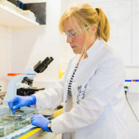
I’m starting this one immediately after having seeded some cells in a snazzy new device designed to allow cell growth in 3D. More specifically, the way I’m using it is to grow cells that make up the blood brain barrier in such a way that they form tubular networks so I can look at things flowing through them, getting stuck in them and leaking out of them. The importance of all of this will shortly become clear because today we’re going to talk about organ-on-chip technologies and organoids and their use in dementia research.
As usual I will start with an apology. This is definitely not my field so the majority of my scant knowledge comes from desperately quizzing my colleague Paul, who has been doing this stuff for years and is exceedingly good at it (Paul Holloway if you want to Google-stalk him). Basically I’ve jumped in to use the technology in a way that I hate people doing in my field so I must be driving him absolutely insane. Irrespective, these techniques are becoming more and more prevalent and they’re useful to know about, in case you feel the need to apply them to your own research so today we’ll cover some history and some of the basics of what they do.
Let’s start with the basics of cell culture. In the very late 1800s, a chap called Claude Bernard did some fairly horrific experiments which proved that individual cells can continue to live after the death of the whole body. Between 1900 and 1950 there were a variety of discoveries that refined what we now know as cell culture – the use of trypsin, the isolation of nutrient profiles for different cells, etc. Of these discoveries the prize for most interesting experiment (at least from the point of view of scientific curiosity), has to go to Alexis Carrel. In his paper On the Permanent Life of Tissues Outside the Organism, he outlines the best methods to start culturing cells for long periods of time, including some principles (such as the continuous changing of medium and the use of 3D substrates) which could even be considered the start of microfluidics. But the fun bit about Carrel is that he later claimed that at least one of the cultures he started during these studies lived for over 20 years, something which nobody has since been able to replicate.
The 1950s also the saw infamous experiments with Henrietta Lacks cervical cancer cells – producing the first ‘immortalized’ cell line. These cells travelled around the world, controversially enabling many discoveries unbeknownst to their donor. Into the 1970s the cancerous elements that allowed dysregulation of the cell cycle were introduced, as well as those regulating telomere length, both of which allowed cells to continuously divide without becoming senescent. Lines such as HEK and Jurkat were developed.
Many of these cells are still used today in research all over the world and they enable essential, fundamental research to take place. But what they fail to recapitulate is the complexity of the whole organism. If we take the vasculature as our organ of choice you can see instantly how culturing cells on a flat surface may not reflect what goes on in the body. For a start blood vessels are tubes, not flat surfaces, but going beyond that there is constant flow of liquid across the surface which not only affects cell growth but also removes waste products. Even Carrel, in his paper from 1912, noted that this might be important. He said that rather than growing cells in a dish, in which they would constantly be sat in their own catabolic products, that rather “The ideal method would have been to give to the cultures an artificial circulation by which they might have secured their nutrition and eliminated their waste products”. Beautiful.
Here is where we bring in the history of microfluidics. From the 1970s onwards, a big drive in the scientific community in general was the miniaturization of things. And between the 70s and the 90s ‘lab-on-a-chip’ became a thing that people were striving for. Whatever you have, make it TINY. For those of you who weren’t around for the birth of the mobile phone revolution, something similar happened there too. Everything folded and became absolutely miniscule, it was marvellous.
In terms of cell culture, the pivotal moment came from the Whitesides group at Harvard, who pioneered the use of PDMS as a substrate. Previous microfluidic chips had been made of glass or other silicates which were great for doing things like isolating specific molecules but weren’t particularly friendly surfaces for growing cells. They not only didn’t grow particularly well on those surfaces, but they also struggled in such tiny spaces, to get enough oxygen and nutrients. PDMS is gas permeable so devices made of this could allow cells to comfortably grow in a normal cell culture incubator. And by using computer aided design software, different channels and different shapes could be produced that allowed cells to grow next to each other, separated by channels of liquid, or separated by other types of barriers.
As you can now see, this opens up worlds of possibility for the growing of cells and this revolution was coined ‘organ-on-chip’. In pulmonary biology, for example, you can grow lung epithelial cells in one channel and pulmonary endothelial cells in the adjacent channel to see how gas exchange might happen at the lung-vessel interface. In the brain you can grow not just endothelial cells, but endothelial cells with astrocytes and pericytes to make a more complete blood brain barrier. You can perfuse your chips to study what happens under flow. You can add neuronal compartments to look at the communication between neurons and the vasculature. If you’re using flow you can introduce air bubbles or fat bubbles to mimic a stroke. You can use iPSCs from patients to see how disease specific mutations affect the formation of vessels or affect communication between cells. The possibilities are almost endless.
As well as trying to recapitulate the cellular complexity by adding all the components back to make an organ on a chip, scientists were also trying to just make cells turn into organs. Which sounds far fetched when you say it like that but organoid research goes back almost as far as cell culture research but breakthroughs didn’t begin to happen until relatively recently.
Some of the first experiments designed to try and create whole organs in a dish used a technique called ‘disaggregation-reaggregation’ which is the cell culture equivalent of taking the clock apart and putting it back together again to see if it still ticks. Needless to say it often did not tick. But these techniques have been perfected and the use of embryonic and pluripotent stem cells has enabled the production of both spheroids and organoids which have a significant degree of cellular complexity. As a rule spheroids are the more simple of the two, where a cell type (or multiple cell types) are grown together but they don’t tend to self-organise into any kind of known structures whereas organoids, which generally use cells with some degree of pluripotency, will do.
In 2013, Lancaster and colleagues published a paper in Nature on cerebral organoids which many referred to as ‘mini brains’. They were aggregated collections of neurons and astrocytes which organised themselves into cortical structures and whose neurons responded to stimulation. Similar organoid systems are now being used around the world to study both brain development and brain disease. The major drawback currently is that these organoids are often not vascularised. As such they have a size limit before the inside of the structure becomes permanently hypoxic, mimicking a sort of ongoing stroke which is potentially not what the people using them really want. But even this hurdle is being overcome by fusing vascular organoids with brain organoids. The authors of the 2022 eLife paper I found on this topic showed that this fusion resulted in the formation of a blood-brain-barrier-like structure and increased neural progenitor cells as well as microglial synaptic pruning.
At the moment I’m mostly just growing endothelial cells on a flat surface and as a consequence feel very out of touch with modern science.
But these techniques are new enough that people are still perfecting them. At some point in the future they will become cheaper and more accessible, they will be less time consuming and more robust. And that’s when I’ll jump in and start using them, when the fashion has gone away and they’re just useable tools for good, solid research. But if you’re out there and thinking about your next project, think about how the cells your interested in talk to all their neighbours, and whether those interactions are an important part of the disease process. And think about whether any of these snazzy new techniques can help you answer any of the questions you’re coming up with. For the questions I’m trying to answer complexity isn’t important just yet, so Paul’s amazing home-made syringe pump and some channel slides will do me for now.

Dr Yvonne Couch
Author
Dr Yvonne Couch is an Alzheimer’s Research UK Fellow at the University of Oxford. Yvonne studies the role of extracellular vesicles and their role in changing the function of the vasculature after stroke, aiming to discover why the prevalence of dementia after stroke is three times higher than the average. It is her passion for problem solving and love of science that drives her, in advancing our knowledge of disease. Yvonne shares her opinions, talks about science and explores different careers topics in her monthly blogs – she does a great job of narrating too.

 Print This Post


