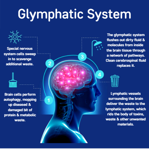
A while ago I read a paper about an interesting idea of how the brain cleared waste from its environment. Ever since I’ve been intrigued to learn more about this phenomenon. So, I thought in this month’s blog I’d give you an insight into the brains waste system – and how it may be important in Alzheimer’s disease (AD). So, saddle up for some fascinating science! So let’s start with the basics, and answer the questions, what is the glymphatic system?
The glymphatic system is a relatively new discovery – only being described in a living system in 2012. In the body the lymphatic system plays an important role in waste clearance, helping to remove excess fluid and waste from the spaces between cells and the blood2. However, lymphatic vessels have only recently been identified in the outer covers of the brain and are still yet to be observed in brain tissue, so how does your brain – which creates an abundance of waste clean itself?
Your brain is surrounded by fluid – known as cerebral spinal fluid (CSF). This fluid is produced inside large ventricles (big holes) inside the middle of your brain. CSF flows through the different ventricles inside your brain into the subarachnoid space (a space which surrounds the brain).
Research indicates that CSF and the fluid that surrounds cells (known as the interstitial fluid) continuously exchange with each other. It’s thought that this exchange occurs due to CSF moving from the subarachnoid space into spaces that lie alongside diving arteries within the brain (these spaces are also known as perivascular spaces). It’s also been suggested that this mixture of CSF and interstitial fluid exit the brain via the spaces surrounding the draining veins of the brain. Once the fluid has exited the brain it is then thought to be reabsorbed into the circulation.
 How was the glymphatic-system discovered?
In the first study to show the glymphatic system in a living system Illif and colleagues imaged the movement of small fluorescent molecules which had been injected into the CSF in mice. When they injected the tracers into the large ventricles in the middle of the brain, they observed little movement of the tracers into the brain tissue. However, when they injected the tracers into CSF in the subarachnoid space (the space that surrounds the brain) the tracers entered the brain tissue!
They suggested that CSF enters the brain and flows in the space alongside arteries that dive down into the brain. These diving arteries have specialised cells called astrocytes which surround them, which help form the blood brain barrier – an important membrane that protects the brain from toxins. Astrocytes also have specialised end feet, which wrap around blood vessels of the brain and within these end feet are specialised channels that permit the movement of water through them.
The authors were curious to understand the role of these specialised water channels in glymphatic clearance. To investigate this, they used a mouse model that had been modified to lack these water channels. Intriguingly, they found that the flow of CSF along the pathway they’d previously discovered was slowed in these mice. Further to this, they also reported a reduction in the clearance of solutes from the fluid that surrounds cells in these mice. From this, they concluded that these water channels, on the end feet of astrocytes are important in the bulk flow of CSF movement into and out of the brain.
How does this waste clearance system link to AD?
There is increasing evidence to suggest that AD may occur due to an imbalance between the production and clearance of proteins within the brain, one of which being Amyloid Beta (AB).
In the study previously mentioned the team also injected soluble AB into the brain to establish how this protein is cleared1. They found that the protein was removed from the brain in the pathways they’d previously proposed. What’s more, AB clearance was reduced in mice which lacked the specialised water channels on the end feet of the astrocytes.
Studies from the same group have also shown that tau, another protein which builds up in the brain in AD can also be cleared from the brain along this same pathway. Further to this, a different research group used a different technique to image the glymphatic pathways in a tau mouse model and found supporting evidence that extracellular tau can be cleared from the brain via the glymphatic pathway. They also reported that in regions of the brain where they found a greater number of tau deposits, that they also found less specialised water channels on the astrocytic end feet. Furthermore, they demonstrated that the application of a drug that inhibits these water channels reduced the exchange of CSF and the fluid that surrounds brain cells, as well as reducing the clearance of tau.
All of this together adds support to the idea that the glymphatic system may be an important waste clearance system of the brain.
Controversies around the glymphatic system
As with all research there are drawbacks and, like most ideas in science there is always some controversy – the glymphatic system is definitely an idea with a lot of controversy surrounding it.
One issue with research investigating the glymphatic system is that most of the studies have been conducted in animals, and very few studies have visualised this system in humans. This is due to a number of reasons, one being that some of the methods currently used in animals, such as two-photon imaging can’t be used in humans.
It has also been proposed that the space surrounding arteries within the brain (perivascular space), where the CSF flows may be altered when brain tissue is removed after death and fixed. Animal studies have shown that the size of the space surrounding arteries in the brain is reduced in fixed tissue. Therefore, some researchers suggest that it’s difficult to fully investigate glymphatic clearance after death. However, perivascular spaces have been observed in humans using MRI.
Finally, it’s important to note that we still do not understand the mechanism of how the specialised water channels aid the movement of CSF. More research into the mechanisms of this are certainly needed.
Overall, it’s clear that this area needs a lot more research for us to fully understand what the glymphatic system is and how it may be important in brain diseases like AD. However, the research so far is interesting and any new avenues that may aid our understanding, as well as serve as potential therapeutic targets for neurodegenerative diseases are worth further investigation.

Beth Eyre
Author
Beth Eyre is a PhD Student at The University of Sheffield, researching Neurovascular and cognitive function in preclinical models of Alzheimer’s disease. Beth has a background in psychology, where she gained her degree from the University of Leeds. Inside and outside the lab, Beth loves sharing her science and we are delighted to have her contributing as a regular blogger with Dementia Researcher, sharing her work and discussing her career.

 Print This Post


